General
![]()
![]()
Domains: Clinical practice, Leadership & accountability
No CPD Points

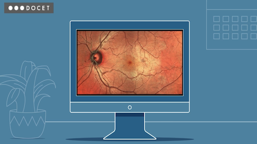
The development of digital imaging systems for optometry has reached the point where they are an integral part of eye health assessment. In this four-part series, we will cover image capture and storage, digital cameras, scanning laser ophthalmoscopy (SLO) and optical coherence tomography (OCT).
Before you start the third part of the series, please ensure that you have either completed Part 1 - Image Capture and Storage and Part 2 - Digital Cameras or you are familiar with the content.
Part three focuses the following:
- SLO instruments, as standalone instruments or incorporated within OCT units
- Various methods of image capture, both central and wide field
- How the use of selected colour channels offers detail of specific clinical tissues and structures and show examples of diagnostic benefits.
To learn how to achieve the best scans with OCT instruments, go to Modern Imaging in Eye Care: Part 4 - OCT.
First published: March 2019
Last reviewed: April 2022