Section

General


No CPD Points


Course Summary

Simon Browning & Susan Cowling (16:21)

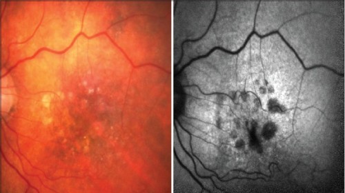
Autofluorescence, the natural emission of light by biological structures, occurs in the retinal pigment epithelium when the fluorophore lipofuscin produced by photoreceptors is stimulated.

A new technology called ‘fundus autofluorescence imaging’ has been developed to allow more accurate detection of autofluorescence in the eye with the aim of identifying retinal diseases when these are not otherwise evident. Here, Susan Cowling asks community optometrist Simon Browning about autofluorescence in the eye, and the implications of the new imaging technology for a wide range of ocular health issues.

First published in Docet OQ88 (2013).

    Open all

    Instructions: Clicking on the section name will show / hide the section.

    View only 'Topic 1'

    • An overview of the scientific basis for each technique.
    • An outline of how the instruments aid examination and diagnosis.
    • An indication of the advantages or potential advantages of each over conventional techniques such as OCT.
    • An indication of which patients might benefit and the advantages for community optometrists.

      View only 'Topic 2'
      View only 'Topic 3'

      References:

      • Kumar N, et al. 2012. Retinal pigment epithelial cell loss assessed by fundus autofluorescence imaging in neovascular age-related macular degeneration. Ophthalmology 120 (2), 334-341.
      • Nandakumar M, Byzney S, Weiter JJ. 2012. Lipofuscin and the principles of fundus autofluorescence: A review. Seminars in Ophthalmology 27 (5-6), 197-201.
      • Moreno JA, et al. 2012. Sustained translation repression by elF2a-P mediates prion neurodegeneration. Nature 485 (7399), 507-11.
        View only 'Topic 4'

        Please give your feedback on this course. Click on the link below to launch the evaluation form.